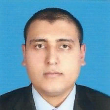
Mohammed Tarek GadAllah
Work place: Department of Industrial Electronics and Control Engineering, Faculty of Electronic Engineering, Menofia University, Menofia, Egypt
E-mail: Mohamed_msc_1986@yahoo.com
Website:
Research Interests: Medical Image Computing, Image Processing, Image Manipulation, Image Compression
Biography
Mohammed Tarek GadAllah was born in Shebeen El-Koom, Menofia Governorate, Egypt on December-29th 1986. He is now a post-graduate master-student at Industrial Electronics and Control Engineering Department, Faculty of Electronic Engineering, Menofia University - Menofia, Egypt. Mohammed was awarded the Degree of Bachelor of Electronic Engineering in July 19 th , 2008 from Industrial Electronics and Control Engineering Department, Faculty of Electronic Engineering, Menofia University - Menofia, Egypt. Mohammed had started learning electronic circuits designing and fabrication under supervision of Dr. Eng. Belal Abozalam at the faculty of electronic engineering, Menofia University, Egypt. He has more than one achievement in designing and fabricating electronic circuits under supervision of Dr. Eng. Samir Badawy. Mohammed, as well as this paper, has Two Published papers [4], [5] accepted & presented in two different conferences. His previous and current researches interests include: Ultrasound Tissue Characterization, Biomedical Image Processing, and Image Processing
Author Articles
Visual Improvement for Hepatic Abscess Sonogram by Segmentation after Curvelet Denoising
By Mohammed Tarek GadAllah Mohammed Mabrouk Sharaf Fahima Aboualmagd Essawy Samir Mohammed Badawy
DOI: https://doi.org/10.5815/ijigsp.2013.07.02, Pub. Date: 8 Jun. 2013
A wise automated method for wisely improving the visualization of hepatic abscess sonogram, a modest trial is being done to denoise and reduce the ultrasound scan speckles wisely and effectively. As an effective way for improving the diagnostic decision; improved sonogram for hepatic abscess is reconstructed by ultrasound scan image segmentation after denoising in Curvelet transform domain. Better sonogram visualization is required for better human interpretation. Speckle noise filtering of medical ultrasound images is needed for enhanced diagnosis. Double thresholding segmentation was applied on, an ultrasound scan image for a Liver with amebic abscess, after it had been denoised in Curvelet transform domain. The result is enhanced wise effect on the hepatic abscess sonogram image's visualization which improves physicians' decisions. Moreover, this method effectively reduces the memory storage size for the image which consequently decreases computation processing time.
[...] Read more.Other Articles
Subscribe to receive issue release notifications and newsletters from MECS Press journals